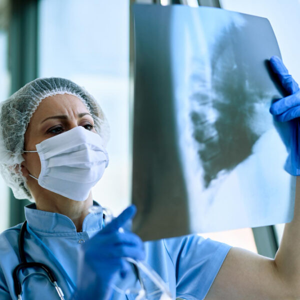
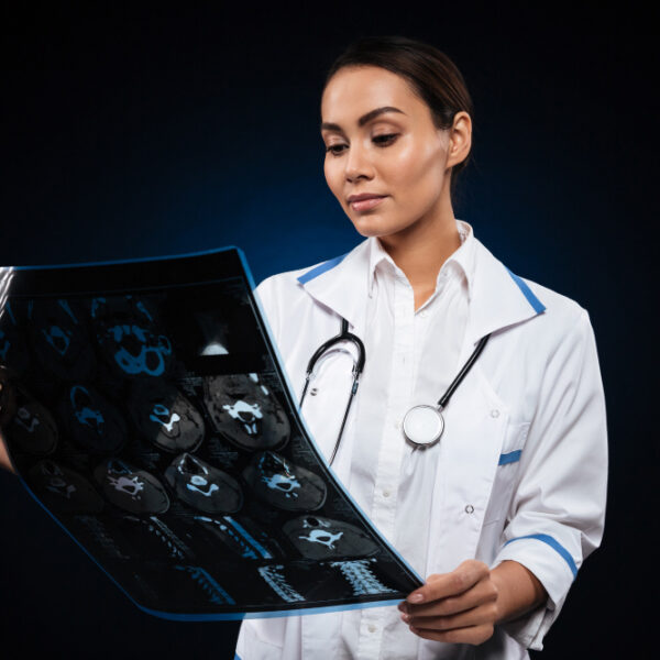
The Department of Radiology offers a wide range of imaging services to assist with diagnostic & therapeutic interventions. The department supports several other specialities across the hospital, including surgical & medical departments. It provides both diagnostic & therapeutic interventional radiology services, using advanced imaging modalities to cater to patient needs. Through this department, the following services are provided and/or accessible.
Investigation services
Besides round-the-clock access to haematology, microbiology & pathology lab services, and conventional radiology services will be provided to support accurate diagnosis. Conventional radiology will form the backbone of the department. Our services includes imaging modalities like computed tomography (CT) & magnetic-resonance imaging (MRI) scans, colour Doppler, digital imaging, digital subtraction angiography (DSA), and digital spot mammography (DSM) with stereotactic breast biopsy. Basic X-rays of the chest, abdomen, and bones will be performed. The department will be equipped with a digital X-ray machine and a mass-miniature radiography (MMR) unit. Image intensifier TV systems will be used for fluoroscopy-guided investigations such as barium studies, intravenous urography, and angiograms. These advanced systems will allow for precise diagnostics and reduced radiation exposure to patients. A 1000-mA X-ray unit with digital radiography & fluoroscopy capabilities will allow for specialised contrast procedures like barium swallow, barium meal, myelograms, and DSA. DSA will be utilised to provide high-quality images of blood vessels, especially in angiography, vascular interventions, and endovascular procedures. The system will be used for diagnosing and treating vascular conditions such as aneurysms, stenoses, and arteriovenous malformations. The DSA system will enable precise imaging of blood vessels with minimal contrast use, making it valuable for vascular assessments, such as in angiography and vascular surgery planning.
The department will offer colour ultrasonography (USG) and colour Doppler for both diagnostic & therapeutic purposes. USG will be used to evaluate abdominal organs, the pelvis, the thyroid, and other structures like breasts, the scrotum, and eyes. Doppler studies will assess blood flow and diagnose vascular abnormalities, including narrowing or blockages of blood vessels. Portable USG will also be available for bedside imaging, including intraoperative use.
The spiral CT scanner will provide advanced imaging of the chest, the abdomen, and other areas, offering dual-phase contrast scans to evaluate tumours and other abnormalities. The CT angiography system will be used for visualising blood vessels, while CT-guided biopsy and bone mineral densitometry will assist in both diagnostic & therapeutic procedures. The department will use echo-speed MRI for highresolution imaging of the brain, spine, musculoskeletal system, and abdominal organs. The MRI system will support advanced techniques like magnetic-resonance angiography (MRA), magnetic-resonance cholangiopancreatography (MRCP), and diffusion imaging to assist in diagnosing conditions such as brain infarctions, tumours, and obstructive jaundice. Functional MRI will be used for brain mapping and presurgical planning. The department will provide functional magneticresonance (MR) studies, including MR spectroscopy, magnetisation transfer imaging, phase imaging, and perfusion imaging.
These advanced techniques will allow for the assessment of brain function, organ perfusion, and the detection of early lesions in various tissues. These imaging techniques complement one another and provide comprehensive diagnostic support. It will support various departments with diagnostic imaging and assist in detecting conditions like tumours, fractures, infections, vascular anomalies, and other pathologies.
Operating theatre (OT) and intervention services
Various interventional & therapeutic procedures are performed to support the treatment of cancer patients and alleviate pain or other symptoms. These procedures include percutaneous nephrostomy, stenting, percutaneous transhepatic biliary drainage, tumour embolisation, and celiac plexus block. Additionally, fine-needle aspiration cytologies (FNACs), or biopsies, are carried out on deep-seated tumours under the guidance of USG, CT or fluoroscopy to obtain samples for cytological or histopathological diagnosis. Interventional radiology includes neurological, vascular & nonvascular intervention procedures.
The following are the neurological intervention procedures.
- Guglielmi detachable coil (GDC) embolisation of intracranial aneurysms
- Carotid angioplasty and carotid stent placement
- Intracranial arteriovenous malformation treatment
- Treatment of vein-of-Galen aneurysm malformation (VGAM)
- Carotid cavernous fistula treatment
- Embolisation of skull-based tumours such as meningioma, nasopharyngeal angiofibroma, and paraganglioma (glomus tumour)
- Cranial-vessel thrombolysis
- Therapeutic & diagnostic carotid occlusion
- Intracranial angioplasty and papaverine infusion for treating spasms
- Petrosal sinus sampling
- Embolisation of scalp arteriovenous malformations
- Embolisation of facial arteriovenous malformations
- Embolisation of spinal arteriovenous malformations
- Embolisation of spinal vascular tumours
- Vertebroplasty for treating vertebral complications caused by osteoporosis, metastasis, multiple myeloma and haemangioma
- Spinal biopsy procedures
- Facet joint steroid injection
The vascular intervention procedures are as follows.
- Balloon angioplasty and stent placement in steno-occlusive lesions in the the aorta, peripheral vessels, and renal arteries
- Embolisation of routine & emergency renal lesions, gastrointestinal lesions, lung lesions, and peripheral malformations
- Stent grafts
- Treatments for aneurysms and pseudoaneurysms
- Treatment for arteriovenous fistula (AVF)
The nonvascular intervention procedures includes the following.
- Image-guided FNACs (biopsies)
- Interventional uroradiology
- Percutaneous nephrostomy
- Antegrade ureteral stent deployment
- Drainage of perinephric collections
- Transrectal prostatic biopsies
- Urethral strictures, dilatation and stents
- Interventional therapy of infertility (Fallopian-tube recanalisation)
- Biliary tract interventions (e.g., percutaneous biliary drainage, complex hilar reconstruction with metallic stents, percutaneous management of postsurgical strictures, percutaneous drainage of postoperative bilomas, gallbladder intervention, cholecystostomy, gallbladder ablation)
- Gastrointestinal interventions (e.g., gastroenteric feeding, gastrostomy, enteral feeding, oesophageal intervention, metallic stents, colonic interventions such as cecostomy and stents)
- Drainage of abdominal abscesses
- Interventional procedures in thorax (e.g., empyema drainage, bronchial stents, and bronchopleurocutaneous fistulae)
- Hepatic interventions (e.g., percutaneous ablation of liver tumours, arterial embolisation of liver tumours, portal-vein embolisation, percutaneous ablation, and drainage of hydatid cysts)
- Interventional therapy for Budd–Chiari syndrome (e.g., inferior vena cava recanalisation, hepatic vein recanalisation, using metallic stents, transjugular intrahepatic portosystemic shunt/TIPS)
Information systems
A radiology information system (RIS) and a picture-archiving and communication system (PACS) will be implemented to support the digital documentation, archiving, transfer, and viewing of imaging data. In the initial phase, digital imaging modalities such as CT, MRI, colour Doppler, and digital radiography units will be interconnected for seamless intradepartmental transfer and storage of images in digital format. Conventional radiographs will be converted to digital formats, with long-term archiving facilitated by PACS using magneto-optical discs and jukeboxes. Additionally, the RIS will be integrated with the hospital information system (HIS), enabling seamless sharing of imaging results across different departments.
Quality control (QC) and radiation monitoring
The department will employ sensitometry and densitometry for quality control of imaging processes. Radiation safety protocols will be followed using thermoluminescent dosimetry to monitor patient exposure during diagnostic & interventional procedures.
Research & development (R&D)
The department will emphasise translational research, aiming to transition findings from laboratory research to clinical applications, thereby enhancing patient care through innovative therapies. The faculty will support research in advanced imaging techniques, exploring areas such as functional MRI, MR spectroscopy, and CT imaging protocols. Research will focus on improving imaging accuracy, minimising radiation exposure, and developing novel noninvasive diagnostic techniques.
Education
Educational initiatives will include regular workshops, advanced training courses, and continuing medical education (CME) sessions. In the future, we plan to introduce the Diplomate of National Board (DNB) course and different paramedical courses in radiology to foster the next generation of specialists. Guest lectures and public symposiums will further support family physicians and promote community awareness of preventive healthcare. These will include hands-on training in radiographic procedures, CT imaging, MRI techniques, and interventional radiology.
Fast Checkup
We providing quality healthcare services to our community
Medical Experts
We providing quality healthcare services to our community
24/7 Services
We providing quality healthcare services to our community